Smooth Muscle Diagram : Smooth Muscle Labelled Diagram - Human Anatomy Body
Smooth Muscle Diagram : Smooth Muscle Labelled Diagram - Human Anatomy Body. Smooth muscle, muscle that shows no cross stripes under microscopic magnification. For example muscles of limbs. It is the main muscle of respiration. These cells have fibers of actin and myosin which run through the cell and are supported by a framework of other proteins. Its wavelike movements propel things through the bodily system, such as food through.
ads/bitcoin1.txt
Smooth muscles are unique in their largely involuntary response, and in their structure. The three types of adrenoceptors present are: Skeletal, smooth and cardiac muscle. The smooth muscle, on the other hand, is found in the wall of blood vessels and viscera (for example in the wall of digestive tract). This smooth muscle can be found surrounding the walls of the blood vessels, the bronchioles in the lungs, and the sphincter muscles used in the gi tract.the gi tract, which is tubular by design, also houses longitudinal muscles in addition to the smooth.
Labeled Cardiac Muscle . Labeled Cardiac Muscle A Labeled ... from i.pinimg.com The term smooth muscle is often used to describe visceral muscle because it has a very smooth, uniform appearance when viewed under a microscope. These are called contact junctions, and they function in much the same way as the skeletal. The smooth muscle, on the other hand, is found in the wall of blood vessels and viscera (for example in the wall of digestive tract). Smooth muscles are unique in their largely involuntary response, and in their structure. , and.the main endogenous agonist of these cell. The three types of adrenoceptors present are: They are spindle shaped and have no striations. Smooth muscle (textus muscularis levis) smooth muscle is a type of tissue found in the walls of hollow organs, such as the intestines, uterus and stomach.
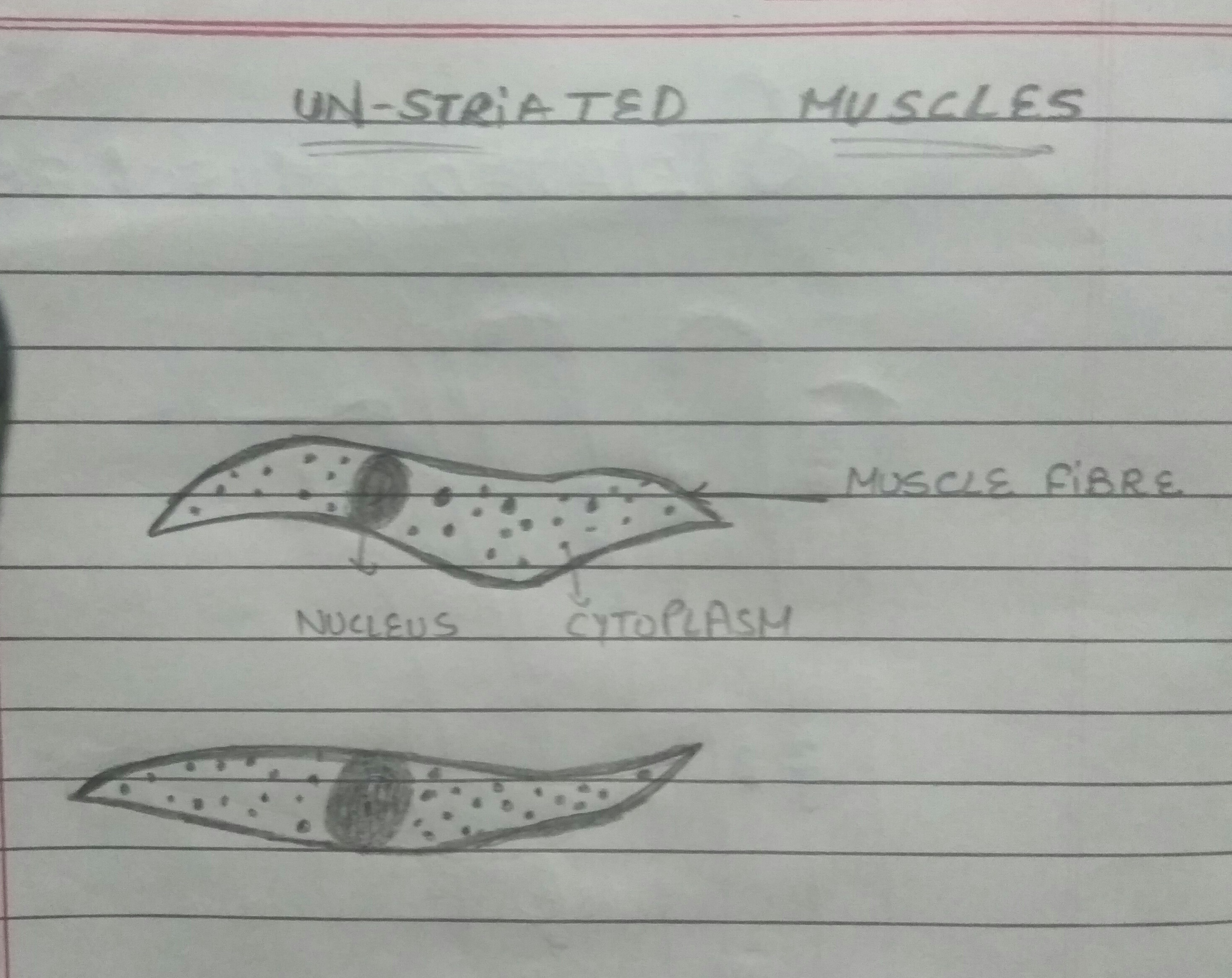
Muscles are made up of highly specialized thin and elongated cells called muscle fibres.the muscle fibres contains specialized cytoplasm called sarcoplasm that contain network of the membrane called sarcoplasmic reticulum.the muscle fibres may be bounded by the cell membrane called sarcolemma.each muscle fibre may contain numerous.
ads/bitcoin2.txt
Skeletal, smooth and cardiac muscle. They are spindle shaped and have no striations. These muscles are often anchored by tendons. Smooth muscle anatomy smooth muscle tissue is also known as visceral muscle tissue. These are called contact junctions, and they function in much the same way as the skeletal. Smooth muscle makes up the walls of hollow organs, respiratory passageways, and blood vessels. Smooth muscle fibers are often found forming sheets of tissue and function in a coordinated fashion due to the presence of gap junctions between the cells. A tendon is simply a fibrous connective tissues, from the muscles to the bone elements. Related posts of smooth muscle diagram muscles of upper back. Smooth muscle tissue, unlike striated muscle, contracts slowly and automatically. The cardiac muscle is only found in the heart wall. These cells have fibers of actin and myosin which run through the cell and are supported by a framework of other proteins. Muscles of upper back 12 photos of the muscles of upper back map of upper back muscles, muscles of the upper back and chest, origin and insertion of upper back muscles, superficial muscles of the upper back, tight muscles of the upper back and neck, human muscles, map of upper back muscles, muscles of the …
They differ in structure and function, for example, in the speed they can contract. Human muscle system, the muscles of the human body that work the skeletal system, that are under voluntary control, and that are concerned with movement, posture, and balance. A ligament is often found in the joints of the body, and are connective fibrous tissues from bone to bone. Smooth muscle anatomy smooth muscle tissue is also known as visceral muscle tissue. It constitutes much of the musculature of
Smooth Muscle Drawing at PaintingValley.com | Explore ... from paintingvalley.com Smooth muscle cells lack the striated banding pattern found in cardiac and skeletal muscle, and they receive neural innervation from the autonomic nervous system. Related posts of smooth muscle diagram muscles of upper back. The smooth muscle, on the other hand, is found in the wall of blood vessels and viscera (for example in the wall of digestive tract). Looking closer at skeletal muscle. Vascular smooth muscle refers to the particular type of smooth muscle found within, and composing the majority of the wall of blood vessels. Smooth muscle is widely distributed in the body. Smooth muscle tissue, unlike striated muscle, contracts slowly and automatically. Muscles of upper back 12 photos of the muscles of upper back map of upper back muscles, muscles of the upper back and chest, origin and insertion of upper back muscles, superficial muscles of the upper back, tight muscles of the upper back and neck, human muscles, map of upper back muscles, muscles of the …
Vascular smooth muscle refers to the particular type of smooth muscle found within, and composing the majority of the wall of blood vessels.
ads/bitcoin2.txt
• smooth muscles respond to stretch only briefly, and then adapts to its new length • the new length however, retains its original _____ seconds or minutes after it has been elongated or shortened (e.g. Muscles of upper back 12 photos of the muscles of upper back map of upper back muscles, muscles of the upper back and chest, origin and insertion of upper back muscles, superficial muscles of the upper back, tight muscles of the upper back and neck, human muscles, map of upper back muscles, muscles of the … Smooth muscle anatomy smooth muscle tissue is also known as visceral muscle tissue. Its wavelike movements propel things through the bodily system, such as food through. Human physiology is a free online course on janux that is open to anyone. Smooth muscle, muscle that shows no cross stripes under microscopic magnification. Smooth muscle makes up the walls of hollow organs, respiratory passageways, and blood vessels. It constitutes much of the musculature of Related posts of smooth muscle diagram muscles of upper back. A ligament is often found in the joints of the body, and are connective fibrous tissues from bone to bone. A tendon is simply a fibrous connective tissues, from the muscles to the bone elements. In skeletal muscle, a single type of somatic nervous system traverses to muscle, where it stimulates organelle in the muscle cells in order to release calcium. Instead, they have bundles of thin and thick filaments.
The smooth muscles perform the functions in the contrast of other types of muscles. Smooth muscles, cardiac muscles and skeletal muscles. It is layered in a distinctive pattern of circular layers. Smooth muscle makes up the walls of hollow organs, respiratory passageways, and blood vessels. Human physiology is a free online course on janux that is open to anyone.
This figure shows smooth muscle contraction. The left ... from oerpub.github.io Diaphragm is also a skeletal muscle. It constitutes much of the musculature of Smooth muscle contracts under certain stimuli as atp is freed. The smooth muscle, on the other hand, is found in the wall of blood vessels and viscera (for example in the wall of digestive tract). The three types of adrenoceptors present are: Broadly considered, human muscle—like the muscles of all vertebrates—is often divided into striated muscle, smooth muscle, and cardiac muscle. Diagram of artery with smooth muscle identification understanding smooth muscles. Muscles are made up of highly specialized thin and elongated cells called muscle fibres.the muscle fibres contains specialized cytoplasm called sarcoplasm that contain network of the membrane called sarcoplasmic reticulum.the muscle fibres may be bounded by the cell membrane called sarcolemma.each muscle fibre may contain numerous.
Smooth muscle anatomy smooth muscle tissue is also known as visceral muscle tissue.
ads/bitcoin2.txt
These are called contact junctions, and they function in much the same way as the skeletal. In this video i have shown the simplest way of drawing muscle drawing. Smooth muscle is a type of muscle tissue which is used by various systems to apply pressure to vessels and organs. Termed unitary smooth muscle or visceral muscle, this type of smooth muscle is the most common observed in the human body, forming the walls of hollow organs. They are spindle shaped and have no striations. In addition, the contractile state of smooth muscle is controlled by hormones, autocrine/paracrine agents, and other local chemical signals. The smooth muscles perform the functions in the contrast of other types of muscles. Human physiology is a free online course on janux that is open to anyone. This smooth muscle can be found surrounding the walls of the blood vessels, the bronchioles in the lungs, and the sphincter muscles used in the gi tract.the gi tract, which is tubular by design, also houses longitudinal muscles in addition to the smooth. Smooth muscle cells are found in the walls of hollow organs, including the stomach, intestines, urinary bladder and uterus, and in the walls of passageways, such as the. Vascular smooth muscle refers to the particular type of smooth muscle found within, and composing the majority of the wall of blood vessels. Compared to skeletal muscle, smooth muscle cells are small. Smooth muscles exhibits a phenomenon called _____ in which:
ads/bitcoin3.txt
ads/bitcoin4.txt
ads/bitcoin5.txt
0 Response to "Smooth Muscle Diagram : Smooth Muscle Labelled Diagram - Human Anatomy Body"


0 Response to "Smooth Muscle Diagram : Smooth Muscle Labelled Diagram - Human Anatomy Body"
Post a Comment